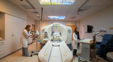
Brigham and Women’s Hospital is the first site in the United States to install a new MRI system designed for safe, high-quality imaging of newborns in a newborn intensive care unit (NICU).
“The installation of this state-of-the-art neonatal MRI system will greatly enhance the Brigham’s research capabilities and elevate and expand neurocritical care for our littlest patients,” says Terrie E. Inder, MD, MBChB, chair of the Department of Pediatric Newborn Medicine. “Putting this technology in the NICU will reduce time and patient risk associated with transporting newborns to a traditional MRI. It will allow MRI access from the first hours of life through the challenging and sometimes life-threatening time in the NICU.”
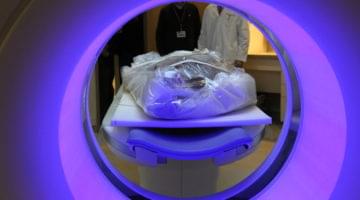
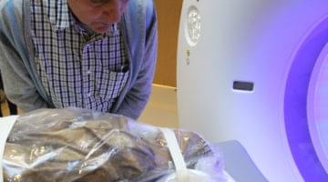
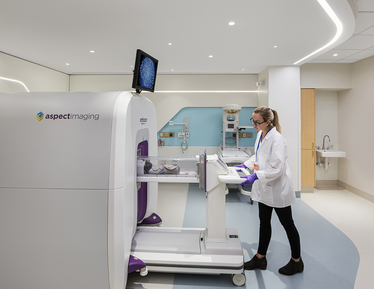
When babies undergo scans, they will be in a temperature-controlled, self-contained incubator bed to minimize movement and provide control of their environment while being continuously monitored for vital signs. Information gained from the MRI can inform the care team and family about any brain injury that has occurred and, in the future, guide which treatments may assist in preventing disability.
Approved by the U.S. Food and Drug Administration in 2017, the neonatal MRI system was built specifically for NICU use. The system is quieter and more compact than traditional whole-body scanners and consumes less power. Initially, it will be used for applied medical research before expanding to clinical use.