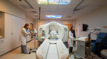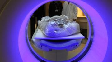
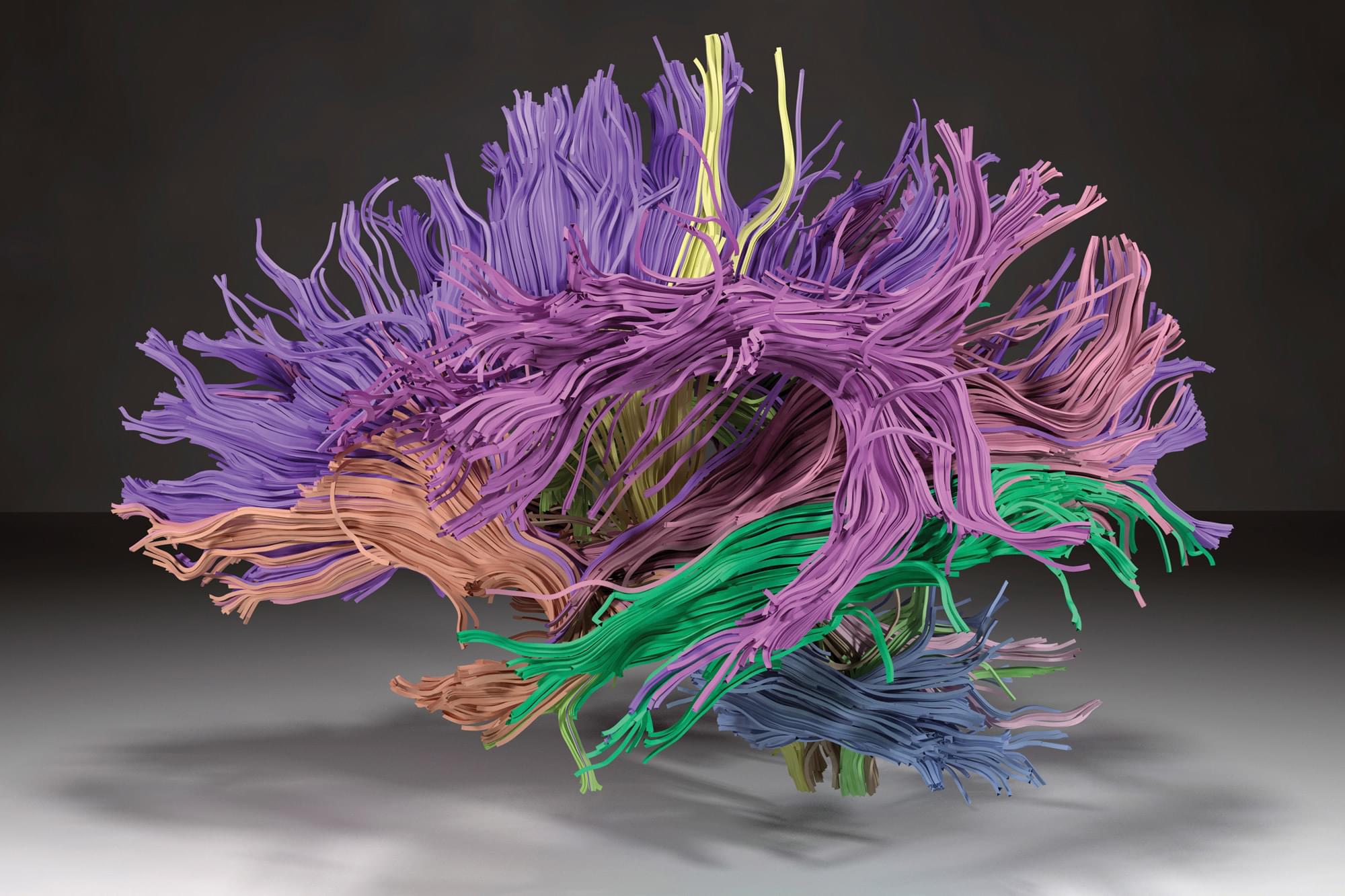
How can scientists see psychiatric disorders in the brain if they are invisible? This 3D model of white-matter fiber bundles illustrates how advances in imaging and machine learning are helping researchers identify deep brain connections that may be crucial to understanding disorders like major depression and schizophrenia.
To create 3D models like this, researchers in the Psychiatry Neuroimaging Laboratory and the Laboratory of Mathematics in Imaging developed an advanced algorithm capable of extracting dMRI (diffusion magnetic resonance imaging) data from thousands of patients’ medical images. The algorithm uses multidimensional imaging data to group together and assign colors to the fiber bundles, resulting in a visualization of brain connections that is remarkably consistent with prior anatomical knowledge. The mapping of these connections can then be used to indicate to researchers whether pathology is present or absent. By measuring differences in the brain’s structure, this mapping tool could reveal important signs of neuropsychiatric disorders, which are not yet detectable through traditional imaging techniques.