
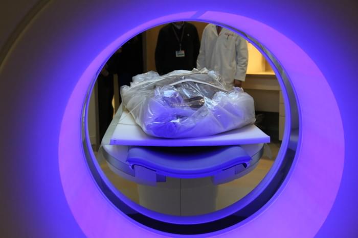
As part of an international research collaboration, Brigham faculty and staff performed CT scans on five mummies from 16th-century Greenland in the Shapiro Cardiovascular Center early last year. The team was looking for plaque in the mummies’ arteries to see if atherosclerosis—a leading cause of death in the U.S. today—was also prevalent centuries ago.
The high-resolution scans of the mummies revealed telltale signs of the disease: hardened calcium deposits in various blood vessels in the chest.
“It’s fascinating to look at humans who lived hundreds of years ago and see if learning about the past could teach us more about the present and future,” says Ron Blankstein, MD, associate director of the Brigham’s Cardiovascular Imaging Program, director of Cardiac Computed Tomography, and a preventive cardiology specialist.
Blankstein was among the experts who performed the CT scans in the Shapiro Cardiovascular Center. He says once the mummies were inside Shapiro, scanning them wasn’t too different from work the cardiovascular imaging team does typically. The mummies were easier to scan than a living patient; normally, the CT scanner must account for the movement of a beating heart.
“It was certainly an exciting and interesting experience,” he adds, “and I hope we can use it to promote awareness of this mostly preventable disease.”